Home » Uncategories » Leg Anatomy Muscles Ligaments And Tendons - Muscles of the Anterior Leg - Attachments - Actions ... - 12 photos of the muscle, tendons and ligaments of leg human.
Tuesday, 22 June 2021
Leg Anatomy Muscles Ligaments And Tendons - Muscles of the Anterior Leg - Attachments - Actions ... - 12 photos of the muscle, tendons and ligaments of leg human.
Leg Anatomy Muscles Ligaments And Tendons - Muscles of the Anterior Leg - Attachments - Actions ... - 12 photos of the muscle, tendons and ligaments of leg human.. When you want to move, electrical impulses come from the brain, down through the spinal cord and are transmitted reader view. Leg anatomy muscles ligaments and tendons : The leg muscles are organized in 3 groups: This important tendon in the back of the calf and ankle connects the plantaris, gastrocnemius, and soleus muscles to. Ligaments, tendons, and muscles play an important role in the function of the hip.
The calf muscles, through the achilles tendon, are the main plantarflexors of the ankle which pulls the foot down. Ligaments and tendons are fibrous bands of connective tissue that attach to bone. Two of these ligaments are in the center of the joint, and they cross each other. Related posts of muscles and tendons of the leg. The human leg, in the general word sense, is the entire lower limb of the human body, including the foot, thigh and even the hip or gluteal region.
Tendons and Ligaments of Ox Leg | ClipArt ETC from etc.usf.edu The tarsal bones are found near the. The achilles tendon is also located in the lower leg. Muscles, either individually or in groups, are supported by fascia. Muscle anatomy diagram 12 photos of the muscle anatomy diagram facial muscle anatomy diagram botox, greys anatomy muscle diagram, groin muscle anatomy diagram, rabbit muscle anatomy diagram, stomach muscle anatomy diagram, human muscles, facial muscle anatomy diagram botox, greys anatomy muscle diagram, groin muscle. The two main calf muscles, gastrocnemius and soleus, run down the back of the calf and join together to form a strong, thick tendon, the achilles tendon, that attaches to the back of the heel. It is a pivotal hinge joint in the leg that allows for a variety of movements (i.e. There are four major ligaments that surround the knee joint. The thigh muscles don't just move your legs.
These muscles allow the ankle to bend downward and outward.
The thigh muscles don't just move your legs. The soft tissue in the knee joint (tendons, ligaments, menisci, cartilage) that provides stability in the knee and hold the bones. Upper leg muscle pain is a very hard pain affect the leg pain as a whole. Unlike tendons, which connect muscle to bone, ligaments connect bones to other bones. The bones consist of organic and inorganic substance. Each of these muscles is a discrete organ constructed of skeletal muscle tissue, blood vessels, tendons, and nerves. The calf muscles (gastrocnemius and soleus), which are connected to the calcaneus via the achilles tendon. Tendons vary in size and are somewhat elastic and attach bones to muscles. These muscles allow the ankle to bend downward and outward. There are up to 13 bursa of various sizes in and around the knee. Muscle and tendon pain in legs, muscles and tendons of the leg and foot. Leg anatomy muscles ligaments and tendons : There are four muscles in the anterior compartment of the leg.
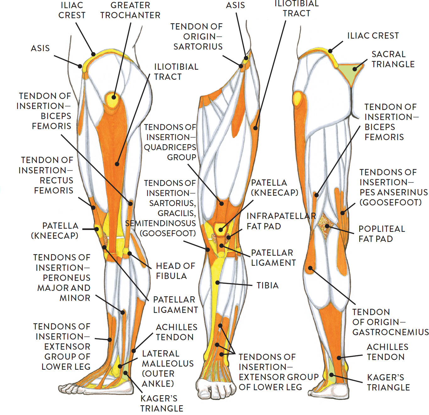
Posterior view of leg showing muscles and tendons involved in ankle movement. Both cross the ankle, but the peroneus longus wraps underneath the cuboid crossing the plantar aspect of the foot as well, and inserts at the base of the first metatarsal. Ligaments, tendons, and muscles play an important role in the function of the hip. Ligaments are structures that connect two bones together. Muscle and tendon pain in legs, muscles and tendons of the leg and foot.
Upper and lower leg, three views from schoolbag.info As with any structure, the human body is built upon a framework that is constructed to carry out a wide range of functions. Two of these ligaments are in the center of the joint, and they cross each other. Major muscles of the ankle. Both cross the ankle, but the peroneus longus wraps underneath the cuboid crossing the plantar aspect of the foot as well, and inserts at the base of the first metatarsal. There are four muscles in the anterior compartment of the leg. The calcaneofibular ligament (cfl), which connects the calcaneus, or heel bone, to the fibula. Muscle anatomy diagram 12 photos of the muscle anatomy diagram facial muscle anatomy diagram botox, greys anatomy muscle diagram, groin muscle anatomy diagram, rabbit muscle anatomy diagram, stomach muscle anatomy diagram, human muscles, facial muscle anatomy diagram botox, greys anatomy muscle diagram, groin muscle. See leg muscle tendon ligament bone stock video clips.
Fibula— a long, thin bone in the lower leg on the lateral side which runs along side the tibia from the knee to the ankle.
These are called the cruciate ligaments and consist of the anterior cruciate ligament and the posterior cruciate ligament. Related posts of muscles and tendons of the leg muscle anatomy diagram. There are up to 13 bursa of various sizes in and around the knee. Master leg and knee anatomy using our topic page. This important tendon in the back of the calf and ankle connects the plantaris, gastrocnemius, and soleus muscles to. Ligaments are structures that connect two bones together. Possibly the most important tendon in terms of mobility is the achilles tendon. Ligaments are soft tissue structures that connect bones to bones. Leg anatomy muscles ligaments and tendons : Anatomy of a knee, tendons, ligaments and common injuries to the knee are described in this article. Tendons and ligaments attach muscles to bones. As with any structure, the human body is built upon a framework that is constructed to carry out a wide range of functions. These fluid filled sacs cushion the joint and reduce friction between muscles, bones, tendons and ligaments.
Related posts of muscles and tendons of the leg muscle anatomy diagram. The system of ligaments in the vertebral column, combined with the tendons and muscles, provides a natural brace to help protect the spine from. This important tendon in the back of the calf and ankle connects the plantaris, gastrocnemius, and soleus muscles to. The achilles tendon is also located in the lower leg. Learn about the muscles, tendons, bones, and ligaments that comprise the knee joint anatomy.
Jumper's Knee (Patella Tendon Overuse Injury, Patella ... from www.thermoskin.com Master leg and knee anatomy using our topic page. The calf muscles (gastrocnemius and soleus), which are connected to the calcaneus via the achilles tendon. The two main calf muscles, gastrocnemius and soleus, run down the back of the calf and join together to form a strong, thick tendon, the achilles tendon, that attaches to the back of the heel. The tarsal bones are found near the. Possibly the most important tendon in terms of mobility is the achilles tendon. Leg anatomy muscles ligaments and tendons. The leg muscles are organized in 3 groups: Muscles, ligaments, & tendons by:
There are four major ligaments that surround the knee joint.
The calcaneofibular ligament (cfl), which connects the calcaneus, or heel bone, to the fibula. The calf muscles (gastrocnemius and soleus), which are connected to the calcaneus via the achilles tendon. Muscle anatomy diagram 12 photos of the muscle anatomy diagram facial muscle anatomy diagram botox, greys anatomy muscle diagram, groin muscle anatomy diagram, rabbit muscle anatomy diagram, stomach muscle anatomy diagram, human muscles, facial muscle anatomy diagram botox, greys anatomy muscle diagram, groin muscle. The gastrocnemius and soleus muscles taper and merge at the base of the calf muscle. Fibula— a long, thin bone in the lower leg on the lateral side which runs along side the tibia from the knee to the ankle. There are many muscles located in the lower leg, but there are three that are particularly well known—the gastrocnemius and the soleus, which are the most powerful muscles in the lower leg, and the anterior tibialis. The bones consist of organic and inorganic substance. Muscles, either individually or in groups, are supported by fascia. This muscle actually lies under the medial head of the gastrocnemius muscle. Anatomy of the knee joints anatomy the knee joint knee tendon lateral knee joint diagram anatomic knee quadriceps muscles knee knee patella femur tibia & fibula. These muscles allow the ankle to bend downward and outward. Ligaments, tendons, and muscles play an important role in the function of the hip. The last of the muscle compartments of the lower leg is the lateral compartment (figure 15) is comprised of two muscles, the peroneus longus and the peroneus brevis.
0 Response to "Leg Anatomy Muscles Ligaments And Tendons - Muscles of the Anterior Leg - Attachments - Actions ... - 12 photos of the muscle, tendons and ligaments of leg human."


0 Response to "Leg Anatomy Muscles Ligaments And Tendons - Muscles of the Anterior Leg - Attachments - Actions ... - 12 photos of the muscle, tendons and ligaments of leg human."
Post a Comment